Which compounds actually target the organelle, which are adjacent, and why the distinction changes assay design.
Compound categories in preclinical literature usually form around a shared mechanism. “Mitochondrial peptides” formed around a shared organelle instead, and the result is a label that now covers several groups of molecules with almost nothing in common beyond the word.
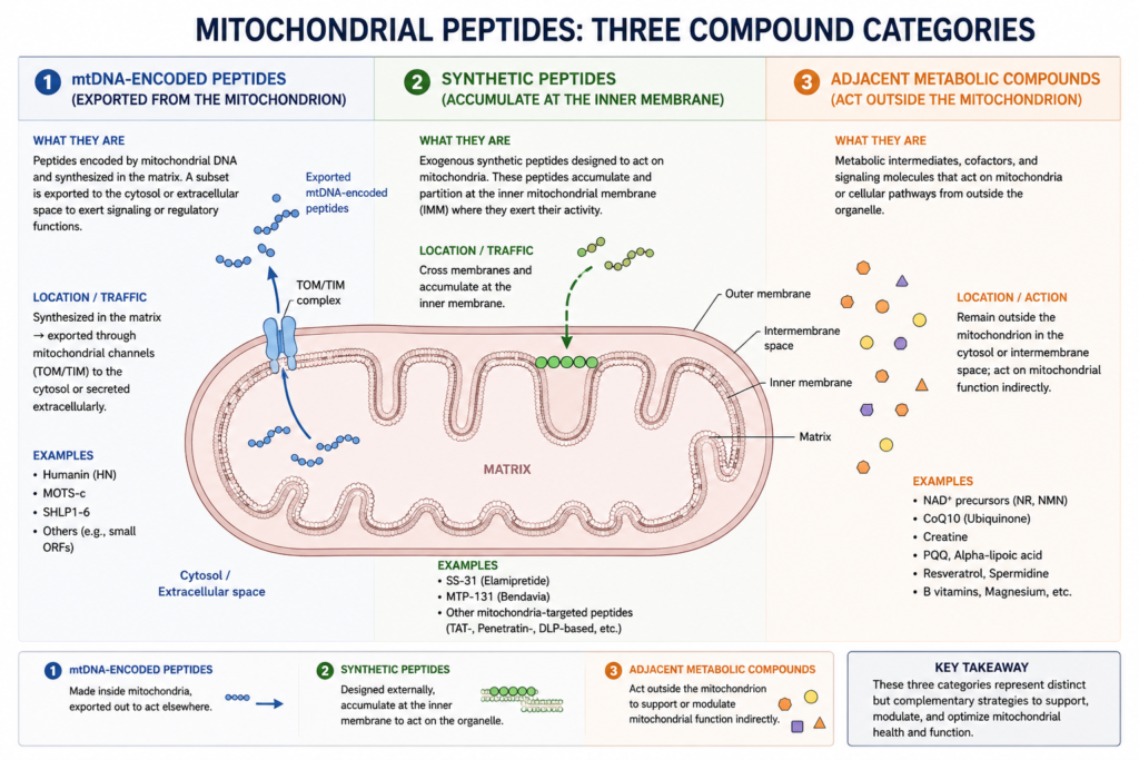
Three distinct things currently travel under that heading. The first is a small family of peptides encoded within mitochondrial DNA that are exported from the organelle to act on nuclear, cytosolic, and systemic targets. The second is a series of synthetic peptides engineered to do the reverse: cross the plasma membrane, accumulate at the inner mitochondrial membrane, and remain there. The third is a loose collection of metabolic compounds, some of them not peptides at all, that appear in the same content because their measured endpoints overlap rather than because they act on the organelle.
The distinction is not a taxonomic nicety. A peptide that signals outward from the mitochondrion and a peptide that acts inside it require incompatible experimental preparations, and a study designed from the category label rather than the mechanism will frequently measure the wrong endpoint and produce a null result that means nothing. This article sets out the three groups, what separates them at the level of receptor and transport, and which assay system each one requires.
Three Categories, One Label
The organising variable is direction of transport relative to the organelle, and it separates the groups cleanly.
Mitochondrial-derived peptides are transcribed from short open reading frames inside the mitochondrial genome. Their transcripts move to the cytoplasm for translation, and the resulting peptides act at destinations outside the organelle that produced them. Their biology is signalling biology, and the organelle is the sender rather than the target.
Mitochondria-targeted synthetic peptides have no endogenous counterpart. They are designed molecules whose defining property is that they cross membranes and concentrate at the inner mitochondrial membrane, where they act on membrane structure and, through it, on bioenergetic function. The organelle is the destination.
The third group is defined by neither of these. It contains endogenous electron carriers, conjugated derivatives of those carriers, transcriptional regulators of mitochondrial biogenesis, and enzyme inhibitors acting on cofactor availability. Some are mitochondrial by location, some by downstream consequence, and none by targeting. Investigators sourcing mitochondria-targeted research compounds will encounter all three groups shelved together, which makes the distinction worth carrying into the protocol rather than leaving at the point of purchase.
mtDNA-Encoded Peptides: Signalling Outward From the Organelle
The mitochondrial genome is small and was long assumed to encode only the thirteen respiratory chain subunits, two ribosomal RNAs, and a set of transfer RNAs. The discovery of short open reading frames nested inside the ribosomal RNA genes established a second class of products, and the peptides they encode are now studied as retrograde signals carrying information from the organelle to the rest of the cell.
MOTS-c
MOTS-c is a 16-residue peptide encoded within a short open reading frame in the 12S ribosomal RNA gene. Its mechanism, characterised by Lee and colleagues in Cell Metabolism (2015), is indirect in a way that distinguishes it from most signalling peptides. The peptide inhibits the folate cycle and the de novo purine biosynthesis pathway tethered to it. Blocking that pathway causes intracellular AICAR, an endogenous AMP-activated protein kinase activator, to accumulate. AMPK activation follows from the substrate accumulation rather than from direct receptor binding.
That upstream position has consequences for how activity is detected. There is no receptor occupancy to measure. The observable chain runs from folate cycle flux to nucleotide pool composition to AMPK phosphorylation state to downstream effectors, and an assay reading any single point in that chain in isolation may miss the signal entirely.
The second mechanistic feature is nuclear translocation. Under metabolic stress, exercise, and aging, MOTS-c moves from the cytoplasm into the nucleus through an AMPK and PGC-1α dependent route, where it associates with transcription factors regulating antioxidant response element containing genes. A review of the retrograde signalling literature describes the resulting arrangement as a feedback loop, since AMPK activation itself promotes the translocation that produces further transcriptional regulation. Skeletal muscle is the primary target tissue identified in the original characterisation, with reported effects on insulin-stimulated glucose uptake and glucose tolerance in diet-induced and age-related insulin resistance models.
Investigators working with MOTS-c and retrograde mitochondrial signalling should note that the nuclear translocation step is the part of the mechanism most easily lost in a simplified preparation, which the assay section below returns to.
Humanin and Its Analogs
Humanin was the first mitochondrial-derived peptide identified, and it established that the mitochondrial genome encodes signalling products at all. It is a 24-residue peptide from the 16S ribosomal RNA region, and its studied activity is cytoprotective rather than metabolic. A 2025 review in Molecular Medicine Reports describes interaction with heat shock protein 90 as one proposed route for that activity, with reported protective effects in cardiomyoblast, dopaminergic neuronal, fibroblast, and retinal pigment epithelium models under oxidative stress conditions, alongside increases in intracellular ATP levels and respiratory rates.
HNG and Colivelin are potency-modified analogs rather than distinct compounds. HNG substitutes glycine at position 14 of the native sequence. Colivelin is a fusion construct joining a humanin analog to activity-dependent neurotrophic factor. Both were developed to raise potency in the neuronal models where native humanin activity is weak, and neither is interchangeable with the native peptide in a comparison.
The SHLP Series
SHLP1 through SHLP6 are six further peptides encoded in the 16S ribosomal RNA region, identified by Cobb and colleagues in Aging (2016). Their characterisation is uneven: SHLP2 and SHLP3 have the most published work, including reported age-related decline in circulating levels, while several members of the series remain largely uncharacterised.
Their relevance to a study design is mostly as a caution. The class is larger than its two well-studied members, the mitochondrial genome may encode further products not yet identified, and any experiment attributing an observed effect to a single mitochondrial-derived peptide is working within an incompletely mapped family.
Synthetic Mitochondria-Targeted Peptides: Accumulation at the Inner Membrane
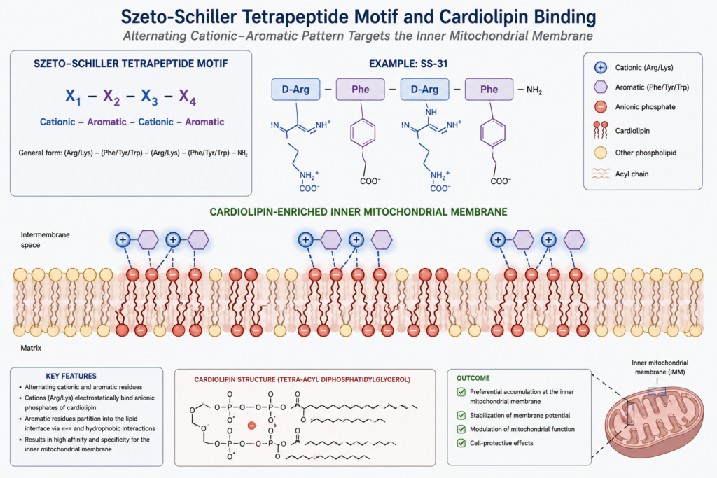
The Szeto-Schiller series is defined by a structural motif rather than by a natural template. As described in a published structure-activity analysis of mitochondria-targeted tetrapeptides, these are synthetic, C-terminally amidated tetrapeptides built on alternating cationic and aromatic side chains.
The transport behaviour that follows from that motif is unusual enough to be the defining property of the class. Despite a formal charge of +3, the peptides traverse the plasma membrane in an energy-independent and non-saturable manner, and then accumulate at the inner mitochondrial membrane at concentrations reported in the range of one thousand to five thousand fold relative to the surrounding compartment. Neither step requires a transporter, and neither saturates, which is why the accumulation gradient is so steep.
SS-31, also known as elamipretide, is the lead compound of the series. The proposed mechanism centres on interaction with cardiolipin, the anionic phospholipid enriched in the inner mitochondrial membrane and required for correct membrane morphology and for the function of membrane-bound respiratory complexes. Binding is understood to stabilise the local membrane environment, and studies with isolated mitochondria and cell cultures report improved electron transfer efficiency, increased ATP production, and reduced electron leak and reactive oxygen species production. Earlier work in cell culture reported protection against induced oxidative stress, with maintenance of membrane potential and reduced lipid peroxidation.
SS-31 also has an approved human use. In September 2025 the United States Food and Drug Administration granted accelerated approval to elamipretide, marketed as Forzinity, for Barth syndrome, a rare inherited disorder in which cardiolipin remodelling is defective, with continued approval contingent on a confirmatory trial. It is not approved in Canada, and the approval concerns a finished pharmaceutical product rather than research-grade material.
SS-02 and SS-20 belong in this section rather than in a list of their own, because their value is comparative. The series was designed to vary the cationic and aromatic residue composition systematically, and SS-20 in particular lacks the dimethyltyrosine residue present in SS-31. Comparing them isolates which structural features drive membrane interaction and which drive the downstream bioenergetic effects, which is a question the lead compound alone cannot answer. A study using SS-31 and cardiolipin binding at the inner mitochondrial membrane as its model system without a structural comparator is measuring the effect without isolating its cause.
Caveat on the cardiolipin mechanism: Cardiolipin binding is the leading hypothesis for how these peptides act, not a settled account. The published structure-activity literature is explicit that several non-mutually-exclusive mechanisms have been proposed to explain the same observed improvements in bioenergetic function, and that progress toward establishing the molecular mechanism of action has come from several independent directions rather than converging on one. Membrane-bound conformation, effects on surface electrostatics, and direct interaction with respiratory complexes have all been advanced. A protocol that treats cardiolipin binding as established fact, rather than as the working hypothesis it is, will interpret an unexpected result as an assay failure when it may be evidence about mechanism.
Prerequisite Condition: The Two Peptide Classes Cannot Share an Assay System
This is where the taxonomy stops being descriptive and starts determining what an experiment can detect.
Isolated mitochondria and permeabilised cell preparations are the standard systems for bioenergetic measurement. They give direct access to respiration rates, membrane potential, and reactive oxygen species production, and they are the appropriate system for a compound whose site of action is the inner membrane. A synthetic targeted tetrapeptide can be evaluated in such a preparation because everything it does happens within the boundary of the preparation.
A mitochondrial-derived peptide cannot. MOTS-c activity depends on folate cycle flux in the cytosol, on nucleotide pool composition, on AMPK phosphorylation state, and on translocation into an intact nucleus. A preparation that has removed the nucleus, permeabilised the plasma membrane, or isolated the organelle has removed most of the mechanism before the compound is added. The peptide will appear inactive, and the null result will be an artefact of the preparation rather than a finding about the compound.
The reverse error is less common but equally available. An intact-cell system with transcriptional readouts is well suited to detecting retrograde signalling and poorly suited to resolving a direct effect on electron transfer efficiency, which will be buried under every other cellular process influencing the same readout.
| Parameter | mtDNA-encoded peptides | Synthetic targeted peptides |
| Genomic origin | Short open reading frames in mitochondrial rRNA genes | None; fully synthetic design |
| Direction of transport | Exported from the organelle | Imported into and retained at the inner membrane |
| Site of action | Cytosol and nucleus | Inner mitochondrial membrane |
| Appropriate preparation | Intact cells with functional nuclear compartment | Isolated mitochondria or permeabilised cells |
| Primary measurable endpoint | AMPK phosphorylation state, ARE-gene transcription, nuclear localisation | Oxygen consumption rate, membrane potential, ROS production |
| What a null result licenses | Nothing, unless translocation capacity was confirmed | Absence of effect on bioenergetic parameters under those conditions |
The final row is the one that most often goes unstated in published methods. A null result is only interpretable if the preparation was capable of detecting a positive one, and if the reagent’s identity and net content were confirmed, and for retrograde-signalling compounds that capability has to be demonstrated rather than assumed.
NAD+ and the Salvage Pathway: Substrate Availability Is Not Targeting
NAD-dependent enzymes, including the sirtuin family, are frequently discussed alongside mitochondrial compounds because sirtuin activity influences mitochondrial biogenesis through PGC-1α deacetylation. The mechanistic relationship is real, and it is also one of substrate supply rather than of targeting.
NAD+ is a cofactor. Raising its availability raises the ceiling on the activity of every enzyme that consumes it, which is a different kind of intervention from a molecule designed to accumulate at a specific membrane. The distinction matters for attribution: an effect observed after raising cofactor availability cannot be assigned to a mitochondrial mechanism without ruling out the other NAD-consuming enzyme families that were equally affected.
Precursor routing adds a second layer. Nicotinamide mononucleotide and nicotinamide riboside enter the salvage pathway at different points and require different enzymatic steps to reach NAD+, so they are not equivalent to each other or to direct NAD+ as a sirtuin pathway substrate in a study where conversion efficiency is a variable rather than an assumption.
5-Amino-1MQ is often grouped here and sits one step further out again. It inhibits nicotinamide N-methyltransferase, the enzyme that methylates nicotinamide and diverts it away from the salvage pathway. Its relationship to NAD+ is therefore competitive rather than contributory, and its relationship to the mitochondrion is downstream of that.

The Adjacent Set: CoQ10, MitoQ, and Transcriptional Regulators
Coenzyme Q10 is the most recognisable compound in this space and a useful entry point, because it illustrates the difference between being mitochondrial by location and being mitochondrial by design. It is an endogenous electron carrier operating between complexes I and II and complex III of the respiratory chain. It is unambiguously part of mitochondrial function. It is not a targeting agent, it has no mechanism for concentrating itself at the inner membrane, and supplying more of it is a substrate intervention of the same general kind as the NAD case above.
MitoQ makes the distinction explicit in its structure. It is a ubiquinone moiety conjugated to a triphenylphosphonium cation, and the conjugation is the targeting mechanism: the lipophilic cation accumulates in response to the membrane potential across the inner membrane, carrying the ubiquinone payload with it. The peptide series described earlier achieves a comparable outcome through an entirely different route, using a cationic and aromatic residue motif rather than a phosphonium cation. Comparing the two is a comparison of targeting chemistries, not of payloads.
SLU-PP-332 belongs to a third mechanism class again. It is an agonist at estrogen-related receptors, which are nuclear receptors regulating the transcriptional programmes governing mitochondrial biogenesis. Its effect, where present, is on the number and composition of mitochondria a cell builds rather than on the function of the organelles already present. That places it on a different timescale from everything else discussed here, since transcriptional and biogenesis endpoints resolve over days rather than the minutes to hours typical of bioenergetic measurement. The exercise-mimetic framing common in secondary coverage describes an intended application rather than a mechanism.
Why Incretin Receptor Agonists Appear in This Category, and Why They Do Not Belong In It
Single, dual, and triple incretin receptor agonists appear routinely in content grouped under mitochondrial function, and the reason is worth stating precisely because it is not a mechanistic one.
These compounds act at plasma membrane G protein coupled receptors. Incretin receptor engagement operates through Gs coupling, adenylyl cyclase activation, and cyclic AMP accumulation, initiating signalling cascades whose immediate effects concern insulin secretion, gastric transit, and central satiety pathways. Glucagon receptor engagement, present in triple agonists and absent from single GLP-1 agents, adds a hepatic component that raises fatty acid oxidation rate and energy expenditure through transcriptional and enzymatic regulation downstream of the same second messenger.
Hepatic fatty acid oxidation occurs in the mitochondrion. That is the entire basis of the association, and it is an endpoint overlap rather than a mechanism overlap. Nothing in the pathway involves a mitochondrial target, mitochondrial accumulation, or any interaction with the organelle that is not mediated by several intervening steps of receptor signalling and gene expression. The distance from receptor to organelle is greater than the distance from a nuclear hormone receptor to the same organelle, and considerably greater than the distance for any compound discussed above.
The practical consequence is that these compounds are not comparators for mitochondrial peptides in a mechanism study. They can share an endpoint with one, which is why they appear in the same metabolic literature, but a design that places a triple GIP, GLP-1, and glucagon receptor agonist reagent alongside a mitochondrial-derived peptide as though the two were acting on the same system has no control for the receptor-level and transcriptional steps that separate them. Grouping by measured endpoint rather than by mechanism is how the category became overloaded in the first place, and this is the clearest example of it.

Frequently Asked Questions
Q1: Why are MOTS-c and SS-31 grouped together when their mechanisms are unrelated?
Direct Answer: The grouping reflects a shared association with the mitochondrion rather than a shared mechanism, since MOTS-c is encoded in mitochondrial DNA and exported to act in the cytosol and nucleus, while SS-31 is a synthetic peptide that accumulates at the inner mitochondrial membrane and acts there.
- Opposite directions: One class carries signals outward from the organelle; the other is transported into it.
- Different sites of action: Nuclear and cytosolic for the derived peptides, inner membrane for the synthetic tetrapeptides.
- Practical consequence: The two cannot share an assay preparation, so a study covering both requires two systems rather than one.
Q2: What determines whether an assay system can detect MOTS-c activity?
Direct Answer: Detection requires an intact cell with a functional nuclear compartment, because the mechanism runs through folate cycle inhibition, AICAR accumulation, AMPK activation, and translocation of the peptide into the nucleus, and a preparation lacking any of those steps cannot produce a positive result.
- Preparations that will not work: Isolated mitochondria and permeabilised cells have removed most of the mechanism before the compound is added.
- Interpreting a null: An absent effect in such a preparation is an artefact of the system rather than evidence about the compound.
- Design implication: Translocation capacity should be demonstrated in the chosen system before a negative result is reported.
Q3: What does cardiolipin binding change about electron transport?
Direct Answer: Cardiolipin is the anionic phospholipid enriched in the inner mitochondrial membrane and required for correct membrane morphology and for the function of membrane-bound respiratory complexes, and the leading hypothesis is that peptide binding stabilises that local environment, with reported effects including improved electron transfer efficiency and reduced electron leak.
- Reported measurements: Studies with isolated mitochondria and cell cultures describe increased ATP production and reduced reactive oxygen species production.
- Mechanistic status: Cardiolipin binding is the leading proposal rather than an established mechanism, and several non-mutually-exclusive alternatives remain under investigation.
- Comparator value: Structural analogs varying the cationic and aromatic residues isolate which features drive the effect.
Q4: How do humanin analogs differ from the native peptide?
Direct Answer: HNG and Colivelin are potency-modified constructs rather than separate compounds, developed because native humanin activity is weak in the neuronal models where the class is most studied, and neither is interchangeable with the native peptide in a comparison.
- HNG: A single residue substitution at position 14 of the native 24-residue sequence.
- Colivelin: A fusion construct joining a humanin analog to activity-dependent neurotrophic factor.
- Attribution problem: Literature describing analog activity does not transfer to the native peptide, and the reverse also holds.
Q5: Why do incretin receptor agonists appear in mitochondrial-function content?
Direct Answer: The association is an endpoint overlap rather than a mechanism overlap, since the glucagon receptor component of multi-agonist compounds raises hepatic fatty acid oxidation, a process occurring in the mitochondrion, without any compound interaction with the organelle itself.
- Actual site of action: Plasma membrane G protein coupled receptors, several signalling steps upstream of any mitochondrial event.
- Why single agonists have a weaker association: Compounds lacking a glucagon receptor component have a correspondingly weaker connection to hepatic oxidative endpoints.
- Design consequence: These compounds are not mechanism comparators for mitochondrial peptides, since nothing controls for the intervening receptor and transcriptional steps.

What the Taxonomy Changes for Study Design
The three groups covered here share a shelf and very little else. Peptides encoded in the mitochondrial genome are signalling molecules that leave the organelle. Synthetic targeted tetrapeptides are engineered to enter it and stay. The adjacent set contains substrate interventions, targeting chemistries built on a different principle, transcriptional regulators operating on biogenesis, and receptor agonists whose connection to the organelle is several steps of signalling removed.
The selection rules that follow are specific rather than general. Studies of mitochondrial-derived peptides require intact cells with functional nuclear compartments and readouts that capture translocation and transcriptional response, and should demonstrate that capability before reporting a null. Studies of synthetic targeted tetrapeptides belong in isolated or permeabilised preparations with respirometry and membrane potential readouts, and gain interpretive power from including a structural analog rather than the lead compound alone. Studies of transcriptional regulators of biogenesis operate on a timescale of days and cannot share a measurement window with either.
Across all three, no shared control arm is available, because the compounds do not share a target. A vehicle control and a positive control drawn from the same mechanism class are the minimum, and cross-class comparison requires that each arm be run in the system appropriate to it rather than in one system chosen for convenience.
The broader point is that a category name is not a mechanism. Where the literature has grouped compounds by the organelle they are associated with, the burden falls on the study design to re-separate them, and the first question in any protocol is not which mitochondrial compound to use but which of these unrelated mechanisms the experiment is actually asking about.
This article is provided for scientific reference in preclinical and in vitro research contexts. It does not describe or recommend any use in humans or animals. Compounds discussed are not approved by Health Canada for any therapeutic indication.
Ready to Advance Your Research?
Browse our catalog of verified research peptides, sourced and shipped exclusively within Canada. COAs available on request.
Disclaimer: All Peptide Wave products are intended strictly for laboratory research use only. Not approved for, nor intended for, human or veterinary consumption.