Reconstitution, Storage, and Why Smaller Vials Protect Your Research
When conducting preclinical evaluation, the integrity of your molecular compounds dictates the accuracy of your data. In the chemical supply industry, maintaining the absolute purity and structural stability of synthesized sequences is a constant challenge. For researchers managing complex laboratory protocols, understanding the exact timeline of peptide breakdown is critical. Improper handling during peptide reconstitution research can easily compromise a compound before it ever reaches an assay.
Using a precise bacteriostatic water reconstitution guide is the first step in protecting fragile amino acid chains from premature degradation. When handling sensitive sequences in BPC-157 research studies, maintaining structural integrity depends heavily on environmental variables like temperature, light, fluid velocity, and container volume. Without strict adherence to thermodynamic and biochemical storage standards, delicate peptide bonds will rapidly fail, introducing unwanted variables into your data.
This educational guide breaks down the specific chemical and physical pathways of peptide degradation. We will analyze the fluid dynamics required during solvent introduction, the kinetics of post-reconstitution decay, and why choosing smaller content vials is one of the most effective ways to mitigate both slow molecular degradation and bacterial contamination risks in the lab.
Chemical and Physical Degradation: How Peptides Break Down Over Time
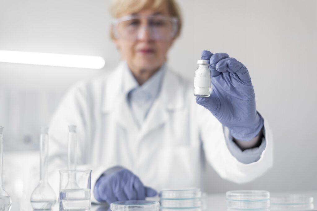
Peptides are short chains of amino acids linked together by covalent amide bonds. While these sequences are structurally distinct from large, folded proteins, they lack the rugged stability of basic small-molecule chemicals. The moment a peptide is synthesized, it is vulnerable to two distinct types of degradation: chemical and physical.
Chemical Breakdown Pathways
Chemical degradation involves the making or breaking of covalent bonds, which completely alters the peptide’s primary structure and transforms it into a completely different, inactive chemical variant.
- Deamidation: This reaction occurs when the side chains of asparagine or glutamine residues lose a functional amine group. This alters the electrical charge of the peptide and twists its shape, preventing it from binding correctly to target receptors in cellular models.
- Oxidation: Exposure to trace amounts of atmospheric oxygen or free radicals primarily damages sulfur-containing amino acids, such as methionine and cysteine. This alters the chemical structure and can completely neutralize the compound’s reactivity.
- Hydrolysis: This is the permanent splitting of the peptide backbone caused by a direct reaction with water molecules. Hydrolysis cuts the amino acid chain into smaller, entirely non-functional fragments.
Physical Breakdown Pathways
Physical degradation changes the secondary or tertiary structure of the molecule without breaking covalent bonds, but it is just as damaging to laboratory accuracy.
- Aggregation: When peptides unfold or lose their native shape, their hydrophobic regions become exposed. To shield themselves from water, these unfolded chains stick to one another. This forms large, insoluble clumps or visible precipitates that ruin the homogeneity of the solution.
- Surface Adsorption: Many peptide sequences are inherently sticky or hydrophobic. They have a natural tendency to cling to the inner glass walls of the vial or plastic laboratory tools, which reduces the actual concentration left dissolved in the liquid.
Reconstitution: The Most Critical Phase
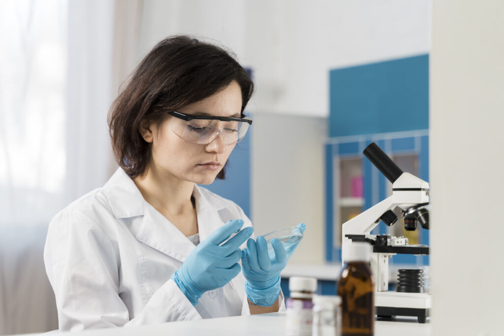
Lyophilization, or freeze-drying, removes moisture from a peptide solution, locking the molecules into a stable, glassy, amorphous powder cake. In this state, chemical reactions are nearly stopped. However, reintroducing a liquid solvent forces the peptide through a high-energy transition phase where the risk of structural damage peaks.
Choosing the Right Liquid
The structural properties of the reconstitution solvent directly dictate the longevity of the liquid peptide. Purified sterile water allows for immediate dissolution, but it leaves the solution highly vulnerable if multiple samples are drawn over time.
The introduction of 0.9% benzyl alcohol in bacteriostatic water serves as a vital antimicrobial preservative. This agent disrupts bacterial cell membranes, preventing the growth of microscopic contaminants during multi-withdrawal laboratory protocols.
However, researchers must note that the specific pH of the solvent can accelerate or decelerate chemical damage; keeping the solution within a peptide’s optimal pH stability window is crucial to avoiding rapid deamidation.
The Fluid Dynamics of Mixing
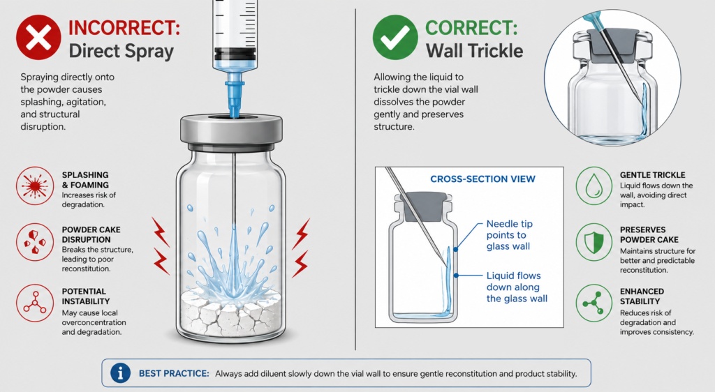
The mechanical force used when introducing liquid into a vial can physically shear fragile peptide structures.
Plunging a solvent straight down onto a lyophilized cake under high pressure creates localized friction and violent kinetic energy. This structural stress can instantly break apart fragile tertiary chains or trigger immediate aggregation.
The correct laboratory method requires angling the syringe needle toward the inside glass wall of the vial, allowing the liquid to trickle down slowly and dissolve the powder cake gently without agitation.
The Post-Reconstitution Clock: Post-Solvent Kinetics
The moment a solvent dissolves the lyophilized cake, a definitive breakdown countdown begins. While dry powder can remain stable for months or years when kept in sub-zero environments, a reconstituted liquid peptide degrades exponentially faster.
Room temperature environments provide the thermal activation energy required to speed up deamidation and hydrolysis. Furthermore, ambient light and ultraviolet (UV) radiation induce photolysis. UV rays hit aromatic amino acids like tryptophan and tyrosine, generating destructive free radicals that cleave the peptide chain apart. Consequently, liquid research peptides must always be shielded from light and kept within strict cold-chain parameters.
Structural Diversity: Why Some Peptides are More Fragile
Not all peptide sequences break down at the same rate. Their vulnerability depends entirely on their length, shape, and amino acid composition.
Short, cyclical peptides or those bound tightly by multiple internal disulfide bridges are highly resilient to mechanical and thermal stress. Conversely, long, straight, linear peptide sequences lack this structural reinforcement, making them highly sensitive to environment-induced unfolding.
When designing laboratory assays, researchers must account for how distinct structural properties alter individual degradation curves:
Fragility Profiles Across Key Research Categories
- Growth hormone secretagogues: Short linear peptides such as ipamorelin and CJC-1295 without DAC lack the disulfide bridges or cyclic structure that stabilise some peptides, so reconstituted solutions should be refrigerated, protected from light and used within the window the supplier’s stability data supports. The receptor pharmacology of this class is covered in growth hormone secretagogues vs exogenous GH.
- Tissue Repair Complexes: While a baseline BPC-157 mechanism of action involves a highly rugged pentadecapeptide sequence, larger molecules like those evaluated in Thymosin Beta-4 research (and its structural fragment, TB-500 mechanism models) possess longer amino acid configurations that are inherently more fragile in solution. Furthermore, combining these distinct sequences into BPC-157 TB-500 combination research vials increases the statistical probability of cross-aggregation, where different peptide chains physically interfere with one another and accelerate clumping.
- Cofactors: NAD+ is not a peptide, and its stability concerns differ. It degrades faster at room temperature and in alkaline solution, so aqueous stocks are usually prepared fresh or stored frozen in single-use aliquots. How NAD+ relates to mitochondrial research compounds is covered in mitochondrial peptides: MOTS-c, SS-31 and how they differ.
Content Vials: Balancing Volume with Protocol Timelines
Because stability curves vary wildly by compound, content configurations must be evaluated through the lens of your specific testing schedule. Stability is entirely peptide and protocol dependent. For instance, one specific compound may be recommended for use within 14 days of reconstitution, while a different, more stable peptide sequence might retain its integrity for 28 days or longer.
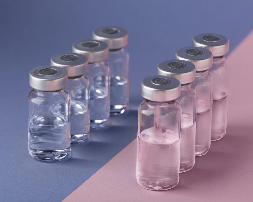
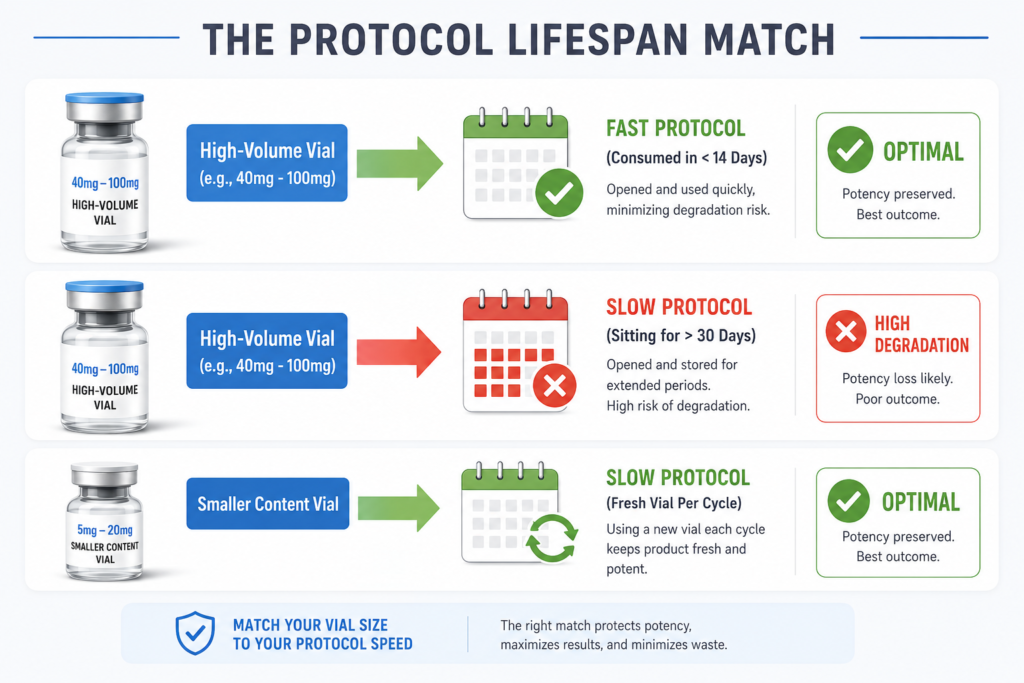
There is no universal “correct” vial size. Certain compounds routinely come in large concentrations like 40mg, 50mg, 100mg, or even 1500mg. If an advanced laboratory protocol using 1500mg vials requires frequent dosing and/or high dosing volumes, the compound may be completely consumed before significant fluid decay occurs.
However, if a protocol calls for a smaller dosing volume over an extended period, a large multi-mg vial will sit in a liquid state for too long. In those specific scenarios, opting for smaller content vials is the better choice. Matching the total vial content to your protocol’s specific lifespan minimizes the risk of structural fragmentation and limits the accumulation of biological impurities caused by repeated needle punctures through the rubber stopper.
Standard Laboratory Storage Summary
To ensure precise, reproducible data, research workflows should always follow these strict storage protocols:
| State | Storage Environment | Target Temperature | Primary Purpose |
| Lyophilized Powder | Deep Laboratory Freezer | -20°C | Long-term structural preservation; prevents moisture accumulation. |
| Reconstituted Liquid | Dedicated Lab Refrigerator | 2°C to 8°C | Slows down hydrolysis and thermal fragmentation post-solvent. |
Contrast dry powder (stable in a freezer at -20°C for months to up to 2 years, depending on the peptide) with mixed liquid (degradation begins within hours or days).
The Bottom Line
Protecting the molecular stability of synthesized compounds requires mitigating a continuous sequence of chemical, physical, and biological threats. Every stage of the handling process serves as a critical inflection point for data integrity. The ultimate precision of your experimental models is directly dictated by variables within the investigator’s control, ranging from the specific physical angle of fluid introduction to the rigid thermal boundaries maintained during long-term storage.
Ultimately, maintaining peak molecular stability comes down to a protocol-dependent awareness. There is no single baseline lifespan for a compound once it enters a liquid state; one sequence may require complete deployment within 14 days, while a structurally reinforced variant might remain viable for 28 days or more. Because stability curves are highly individual, selecting the right compound configuration requires matching the total vial volume to the velocity of your specific testing sequence.
When a protocol calls for rapid, high-volume utilization, a large 40mg, 50mg, or 100mg vial is perfectly optimal, as the sequence is completely consumed before clock-driven fluid decay can compromise the data. Conversely, for extended testing timelines with lower volume requirements, utilizing smaller content configurations ensures fresh, unfragmented sequences are introduced for each new cycle. Aligning the physical container volume with the exact operational window of the protocol is the single most effective way to eliminate data bottlenecks. This deliberate matching guarantees peak purity, minimizes physical rubber stopper wear from repeated needle punctures, and ensures maximum reproducibility for every single evaluation.
Ready to Advance Your Research?
Browse our catalog of verified research peptides, sourced and shipped exclusively within Canada. COAs available on request.
Disclaimer: All Peptide Wave products are intended strictly for laboratory research use only. Not approved for, nor intended for, human or veterinary consumption.